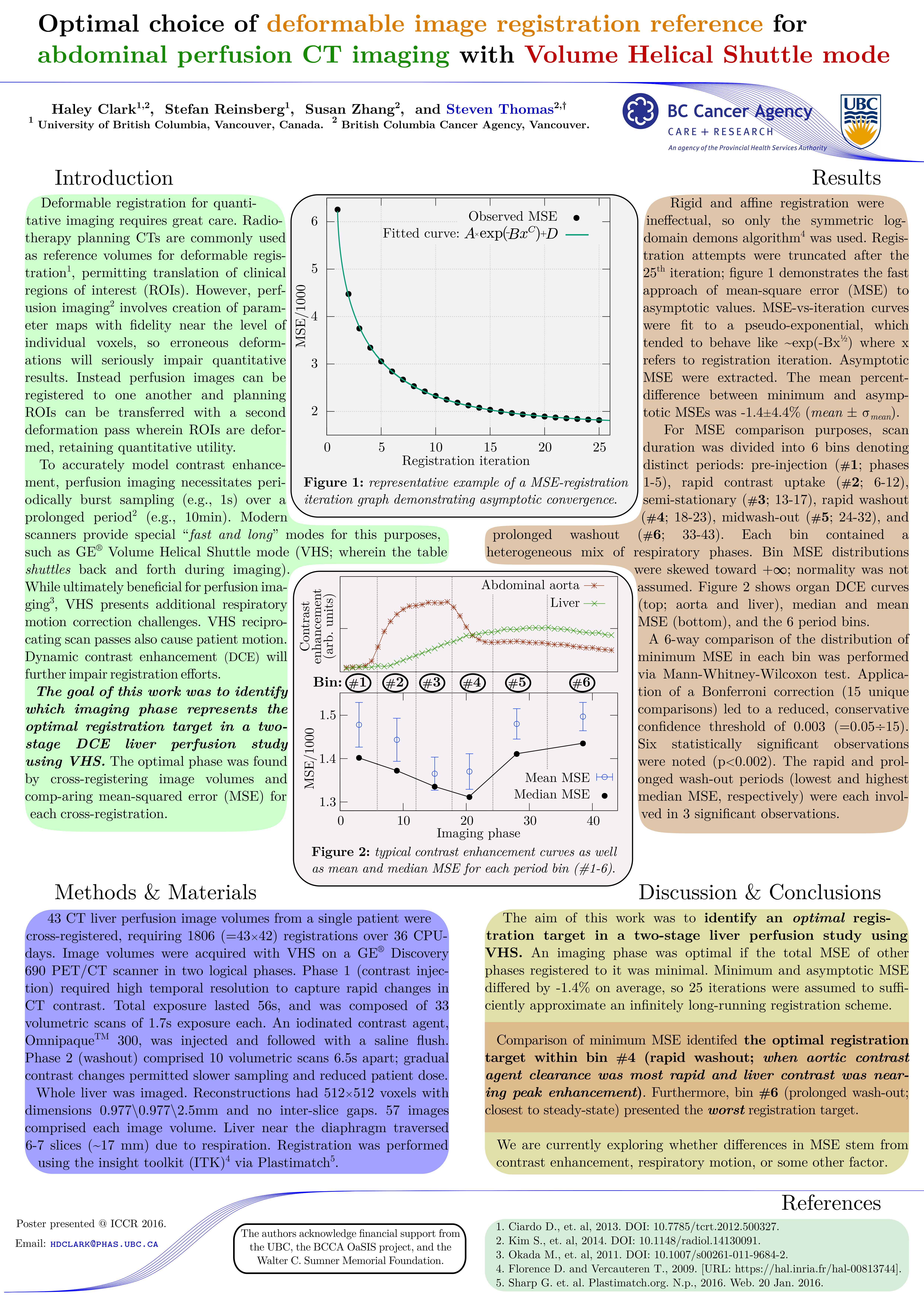
When performing image analysis on a 4DCT or any other volumetric time series (e.g., MRI), patient organ motion can present a significant problem. One approach is to contour all time points separately and analyze each region-of-interest or organ for each time point. This works well for map-reduce style problems, but what if you need to track lobes, compartments, or even individual voxels? In these circumstances it is not uncommon to register contour-to-contour to find a spatial mapping for each time point. This works, but is tedious – why not just deformably register the images together collectively before contouring? Then you only have to contour once, you can track organs, lobes, compartments, and indeed even individual voxels throughout the entire time course if the registration model, nature of the motion, and image site are appropriate.
One problem is that if you have N ‘frames’, it isn’t clear which frame should be selected as the static frame. I investigated this question in the context of liver CT perfusion imaging, where a contrast enhancement solution is administered and accurate tissue tracking are needed for blood flow assessment. My findings were that selection of a frame midway through the series, shortly after arterial enhancement had peaked, provided the best results with the smallest possible deformations. Furthermore, selecting frames from the end of the time series, after both arterial and venous enhancement had peaked, provided some of the worst results.
The following poster provides more rigor and explains my methodology. It was presented along with the video below. Note that this poster is licensed cc-by-nc-sa-3.0.
This poster (abstract) can be cited as:
H. Clark, S. Reinsberg, S. Zhang, and S. Thomas.
Optimal choice of deformable image registration reference for abdominal perfusion CT imaging with Volume Helical Shuttle mode.
Poster presentation at 18th International Conference on the use of Computers in Radiation Therapy, London, UK. June 27→30, 2016.
The following video shows the technique in action for a liver perfusion scan. The original time series is on the left and the corrected series is on the right. A significant reduction in organ motion can be observed, though further work may be needed if voxel-to-voxel mapping is needed.